Live Blood Test & Blood Microscopy
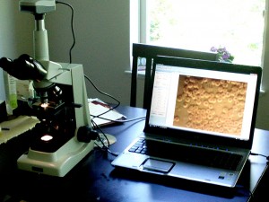
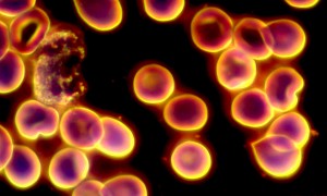
 Live blood cell analysis is used for nutritional and oxidative status evaluations. It is made on a specifically designed powerful digital microscope where light is reflected by blood constituents, what can be seeing in microscope binoculars and image is transferred via camera on the screen of computer, so clear video image appears on the screen. During live blood analysis the main attention is paid not on a particular count of blood constituents but on their shapes and activity. As an old saying says: ‘it is better one time to see, then many times to hear’, this is why live blood analysis when is watched on the screen can be a very good motivating factor as well as showing progress or regress.
French scientist Gaston Naessen in 1946 noted some unusual particles in blood samples he was studying. Because they appear only at 12 noon in May, June and July when the sun light spectrum is very powerful, he developed a different type of microscope, which he called ‘the somatoscope’. Literally he developed a special type of ‘dark field’ microscope in which one could see the interior of living cells in their live state. Using this powerful ‘dark field’ he called particles ‘somatids’ – small bodies. Gaston Naessen discovered that somatids were capable to pleomorphism, and this mean that they could change their shape and their size. They could change from particles to the size of viruses to bacteria, yeast and fungal forms depending on body homeostasis, metabolism and pH of body fluids.
 Blood is the essence of life. It carries our heritage in each strand of DNA. Not only our heritage is transported in every cell in the body, but our blood continuously carries necessary oxygen and vital nutrients to our organs, muscles, bone marrow, and brain. Nutritional deficiencies are also exposed in live and dry blood microscopy. When the cells lack essential nutrients, their structure is impaired and their ability to function is reduced. Most interesting in the observation of live blood is the consistent increase of electrical frequency in and around cells after a person continues on balanced nutrition. This phenomenon is significant since electrical frequency and electrical shield prevents fre radicals from killing healthy red blood cells.
Live and dry blood microcopy is a way of monitoring general health and it is particularly useful in tracking a person’s response over time to the treatments.
How live and dry blood microscopy is done?
In live and dry blood microscopy a drop of blood from a fingertip is placed on a slide under a glass cover slip and is examined under dark field powerful microscope.
What can be evaluated in live and dry blood microscopy?
• Blood coagulation level
• Activity of platelets (thrombocytes)
• Disorders of protein, fat and uric acid metabolism, nutritional status
• Amount and viscosity of red blood cells (RBC)
• Difference in forms and shapes of RBC and terrain
• Types, amount and activity of white blood cells (leukocytes)
• Presence of somatids and their forms
• Speed of blood flow which depends on pH of body fluids, electrical charge, and metabolism
• Oxidative stress
• Influence of electromagnetic field (EMF) on live blood sample exposed to the radiation effect from mobile phone and computer.
Live and dry blood microscopy is used by many famous institutes and clinics all over the world. Many of them use this test for more than 30 years to choose right treatment for their patients, access and monitor progress during treatment, indentify many other things. One of the most scientifically based centres that uses this test is Hippocrates Institute in America (www.hippocratesinst.org).
Hippocrates institute helped many people to obtain health that they lost; they work successfully with different type of cancer, autoimmune disorders, rheumatoid arthritis, adrenal exhaustion and list goes on. They use live and dry blood microscopy for every single guest before and after their treatment, and they say that test does show the difference.
It is a very easy and quick way to see what is happening under the microscope in a tiny drop of blood before and after changing diet or lifestyle.